

Key Takeaways
Why Skin Cancer Detection Matters
Skin cancer is the most commonly diagnosed cancer in the United States, according to the American Academy of Dermatology. The three main types — basal cell carcinoma, squamous cell carcinoma, and melanoma — vary significantly in their growth rates, spread potential, and treatment complexity. Basal and squamous cell carcinomas are far more common and, when caught early, are usually highly treatable. Melanoma is less common but more likely to spread to other parts of the body if not detected at an early stage.
What makes skin cancer particularly well-suited to preventive screening is that it is, quite literally, visible. Unlike cancers that develop internally and may be asymptomatic for years, changes in the skin's surface can often be detected by both trained clinicians and attentive patients. This visibility makes regular self-exams and professional screenings among the most straightforward preventive tools available to most people. For a broader look at how skin cancer checks fit alongside other recommended screenings, see our recommended health screenings by age and sex.
What you will need
How to Perform a Skin Self-Exam and See a Dermatologist
The steps below walk you through assessing your own skin monthly and preparing for professional evaluation. Use these as a practical framework — they are not a substitute for a dermatologist's clinical judgment.
Full-length and handheld mirrors
Allow complete visualization of all skin surfaces, including the scalp, back, and soles of the feet, during a self-exam.
Smartphone camera
Used to photograph spots and compare images over time to detect gradual changes in size, shape, or color.
Bright incandescent or daylight-spectrum lamp
Provides adequate illumination to distinguish subtle color variations and surface texture changes in lesions.
Learn the ABCDE criteria before you start
Dermatologists use a standardized framework called the ABCDEs to assess moles and lesions. Familiarize yourself with each criterion:
- Asymmetry: One half of the mole does not match the other.
- Border: Edges are irregular, ragged, notched, or blurred.
- Color: The mole contains multiple shades — brown, black, red, white, or blue.
- Diameter: The lesion is larger than 6 mm (roughly the size of a pencil eraser), though melanomas can be smaller.
- Evolving: Any change in size, shape, color, or new symptoms such as itching or bleeding.
The "E" — evolving — is considered by many dermatologists to be the most clinically important single indicator, since change over time is a red flag regardless of other features.
Conduct a systematic full-body self-exam
Skin cancers can appear anywhere — including areas rarely exposed to the sun, such as the soles of the feet, between the toes, and under the fingernails. Follow a consistent head-to-toe sequence so you don't miss any area:
- Examine your face, ears, and scalp (use a comb or hair dryer to part hair; a partner or stylist can help).
- Inspect your neck, chest, and torso, including under the breasts.
- Use the hand mirror to view your back, shoulders, and the back of your neck.
- Check both arms, including the underarms, elbows, and between the fingers.
- Examine your lower body: abdomen, hips, groin, legs, and the soles and tops of your feet.
Document any spots you want to track using photographs with a ruler or coin in the frame for scale reference.
Assess your personal risk level
Not everyone carries the same baseline risk for skin cancer. Understanding your risk helps determine how often you should see a dermatologist professionally. Higher-risk characteristics include:
- Fair skin, light hair, or light eyes (lower melanin offers less natural UV protection)
- A history of frequent sunburns, especially blistering burns in childhood or adolescence
- Significant cumulative sun exposure or indoor tanning use
- A personal or family history of melanoma or other skin cancers
- A large number of moles (more than 50) or a history of atypical (dysplastic) moles
- A weakened immune system due to medication or medical conditions
People with one or more of these factors may benefit from more frequent professional screenings. Discuss an individualized schedule with a dermatologist or your primary care provider. For context on how skin checks fit within broader preventive care, see our guide to recommended health screenings by age and sex.
Schedule and prepare for a professional skin exam
A full-body skin exam by a board-certified dermatologist typically takes 15–30 minutes. To make the most of the appointment:
- Arrive with clean, product-free skin and without nail polish, which can obscure nail-bed lesions.
- Bring a list of spots that concern you or that you've noticed changing.
- Be prepared to share your medical history, including any past skin cancer diagnoses and your family history.
- Ask your dermatologist to explain anything they examine — understanding what they are looking for helps you participate in your own care.
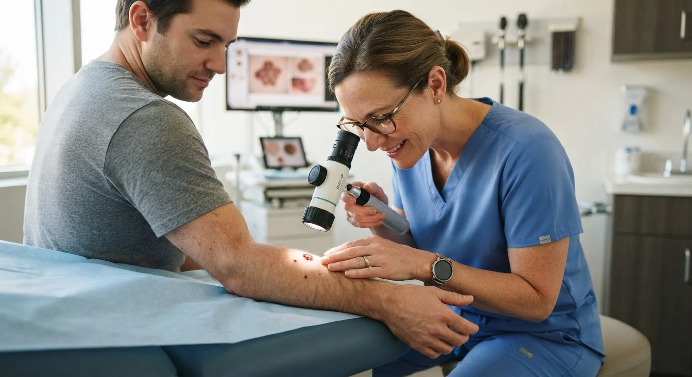
During the exam, your dermatologist may use a dermoscope — a handheld device that uses magnification and polarized light — to evaluate lesions in greater detail than the naked eye allows. If a lesion looks suspicious, they may recommend a biopsy, in which a small tissue sample is removed and examined under a microscope to confirm or rule out cancer.
Establish an ongoing screening routine
Skin cancer prevention and detection are ongoing commitments, not one-time events. General guidance for most average-risk adults includes a professional full-body skin exam at least once a year, combined with monthly self-exams. However, frequency should be individualized based on your risk profile — a conversation with your dermatologist is the best way to arrive at the right schedule for you.
Complement your screening routine with consistent sun protection habits: broad-spectrum SPF 30+ sunscreen applied daily, protective clothing, and avoiding peak UV hours. These habits reduce new damage over time. You may also find it useful to read about common misconceptions that lead people to skip preventive screenings — many of those myths apply equally to skin checks.
Make Self-Exams a Monthly Habit
Choose a consistent date — such as the first day of each month — and examine your skin in a well-lit room using a full-length mirror plus a hand mirror for hard-to-see areas. Take photos of spots you want to monitor over time; side-by-side comparisons are one of the most practical ways to detect gradual changes.
Self-Exams Do Not Replace Professional Screenings
Monthly self-checks are a valuable habit, but they are not a substitute for a trained clinician's eye. Dermatologists use tools and training that allow them to evaluate features invisible to the naked eye. If you have elevated risk factors, discuss an appropriate screening schedule with your healthcare provider.
Seek Care Promptly for Alarming Changes
Do not wait for a scheduled annual visit if a mole or skin lesion changes rapidly in size, shape, or color, bleeds without injury, or is new and growing. Contact a dermatologist or your primary care provider as soon as possible. Early detection meaningfully improves outcomes for most types of skin cancer, and timely evaluation is always appropriate when something concerns you.
This article is for general informational and educational purposes only and does not constitute medical advice. Skin cancer risk and appropriate screening schedules vary by individual. Always consult a qualified healthcare provider or board-certified dermatologist for guidance specific to your health history and circumstances.